
Lesson
Objective:
- Getting to know the structure of a microscope
- Learning how to use a microscope
- View the onion husk cell under the microscope
Teacher’s Guide
- Before proceeding with the lab work, review the safety rules by following the link:
- Divide the students so that there are three students in each group.
- To download the worksheet, go to: worksheet
Theoretical part
Procedure for working with a light microscope
Working with a microscope can be very interesting and exciting, but it requires precision and care.
The first thing to do before working with a light microscope is to check that all parts of the instrument are in place and in good condition. Check that the objective lens and eyepiece are clean and can be wiped down with a soft cloth. Also check that the light is on and working.
Next, select the object you want to examine. It should be clean and dry. The object can be placed on the microscope stage and secured with clamps. Position the stage so that the object is under the objective.
After you have prepared the object, you can start working with the microscope. Turn on the microscope and set the lens to minimum magnification. In this case, the lens should be at a distance of about 1-2 cm from the object. The adjustment can be done with the focus.
After you have set the lens to minimum magnification, you can begin to magnify the object. To do this, turn the knob on the microscope, which is responsible for magnification. Remember to make sure that the object stays in focus.
When you reach the desired magnification, you can begin to examine the object. To do this, you can move the microscope stage with the focus to see different parts of the object. If the object is too dark, you can turn on additional illumination.
At the end of your work with the microscope, you should turn it off and gently remove the object. Be sure to wipe the objective lens and eyepiece with a soft cloth to get rid of fingerprints and dust.
Practical part
Step 1.
Become fully acquainted with the microscope structure and the necessary materials.

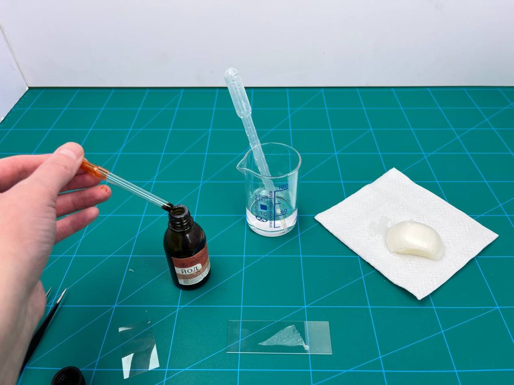
Step 2.
Make sure your slide glass is clean. If it is not clean, it is better to wipe it first. Pipette 1-2 drops of water onto the slide.

Step 3.
Prepare the micropreparation. Remove the inner film from the onion and place it in a beaker.

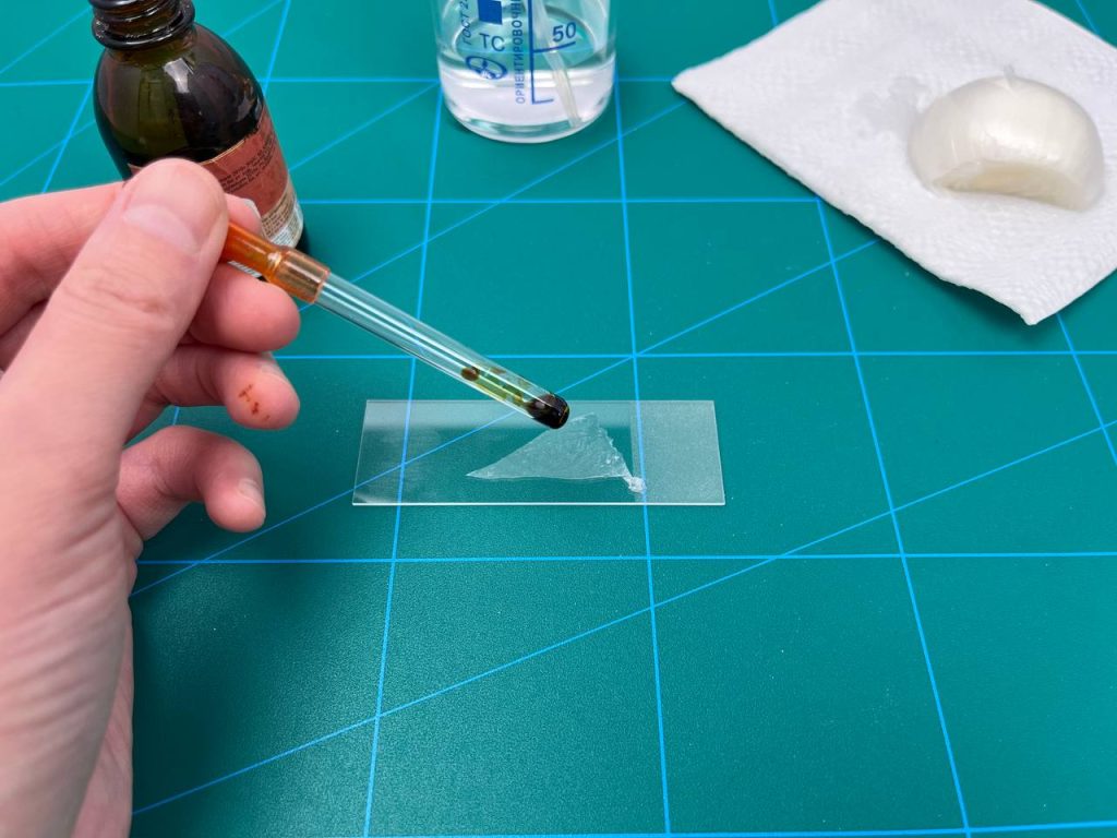
Step 4.
Arrange the onion husks by spreading them correctly on the glass. If there is air remaining in the water in the middle of the onion husks and the glass, remove it. Remove it with a paper towel or filter paper if there is excess water over the husk.
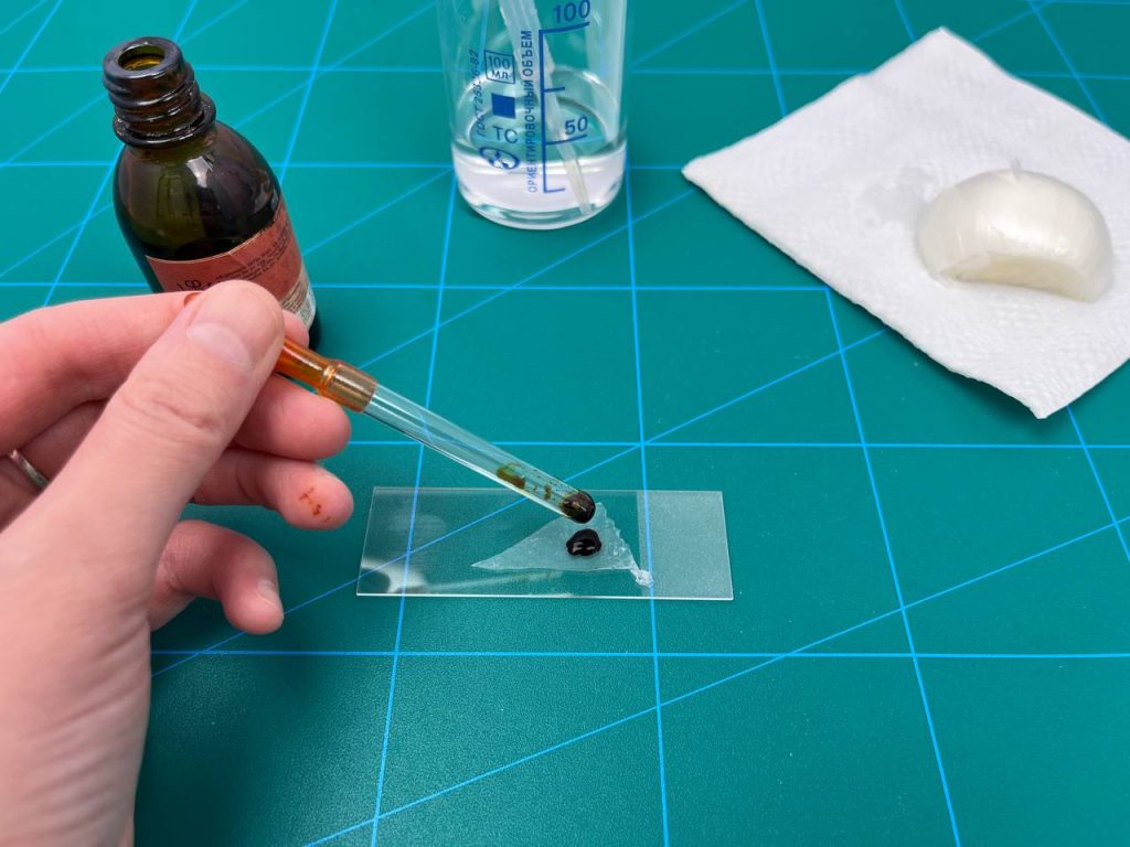
Step 5.
Using a pipette, add iodine to the onion husks. Iodine is used to stain the cells and improve visibility under a microscope. Cover the husk with thick glass.
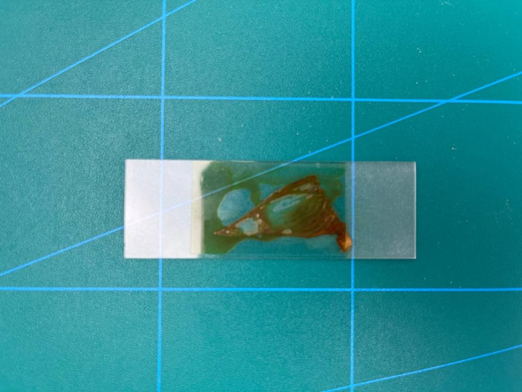
Step 6.
Place the stained specimen under the microscope. Direct the light through the mirror into the opening of the slide where you place the specimen.
Step 7.
Looking through the eyepiece, slowly move the tube through the screws until the image of the object is clear.
Step 8.
Look at the preparation under magnification. Look at the magnification in the eyepiece and the magnification of the lens. You can see the magnification of the preparation in different ways by changing the objective magnification in different ways.
The microscope eyepiece in the image is x16 and the lens magnification is x4, x10 and x40. Accordingly, the magnification is increased 64 times for x16 and x4.

Step 9.
Look at the preparation with the magnification. Screw on the next lens magnification size. The magnification is increased 160 times for x16 and x10.

Step 10.
Look at the preparation with the magnification. Screw on the next lens magnification size. The magnification is increased to 640x for x16 and x40.

Step 11. Make conclusions by answering the questions:
- What is micropreparation and why is it used in the study of the microcosm?
- How do you prepare a micropreparation of an onion husk?
- What tools and equipment would be needed to examine a micropreparation under a beam microscope?
- What structures and cells can be detected when examining a micropreparation of onion husks under an onion microscope?
Conclusion
By doing this practical work, students have developed skills in working with an onion microscope. By studying the peel of an onion, they became familiar with the structure of the cells. They were able to examine the hidden parts and structure of onion peels, invisible to the naked eye, through a microscope lens that could magnify at different levels.
Through working with a microscope, students learned how to set up and use a microscope, know the basic types of microscopes and how to use them, work with preparations, and determine their structure and composition.