
Lesson

11th grade. Biology. Lab #2 Description of the main components of cells using microphotographs
Purpose of the work:
- To study the main components of cells using microphotographs
Expected results:
After completing the work, students can:
- develop teamwork skills
- be able to analyze and summarize the information received
- draw logical conclusions
Teacher’s Guide:
- The task is performed in groups of 3-4 people
- Before starting laboratory work, please read the safety rules by following the link:
- To download the worksheet, follow the link:
Theory
The cell is the basic structural and functional unit of living organisms. All living beings are made of cells, and it is within the cell that life-sustaining processes occur.
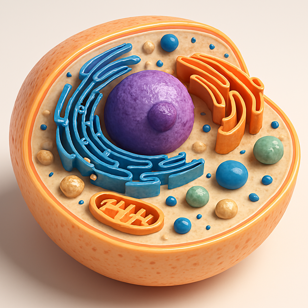
The cell consists of three main components:
- Plasma membrane — separates the cell from the external environment and regulates the exchange of substances. It is semipermeable: allows only necessary molecules to pass inside and removes waste. Receptor proteins in the membrane are involved in signal transmission.
- Cytoplasm — a semi-fluid medium in which organelles are located. Most biochemical reactions occur here.
- Nucleus — stores genetic information in DNA molecules and controls all cellular processes.
Main organelles of the cell and their functions
- Nucleus
The “control center” of the cell. Contains chromosomes made of DNA and proteins. Stores hereditary information that determines the structure and function of the organism. Regulates division, growth, and metabolism. - Nucleolus
Located inside the nucleus. Its main function is the synthesis of ribosomal RNA and the assembly of ribosomal subunits, which later move into the cytoplasm. - Ribosomes
Small non-membrane organelles composed of RNA and proteins. They may float freely in the cytoplasm or be attached to the rough ER. Function — protein synthesis. - Endoplasmic Reticulum (ER)
A network of membrane-bound tubules and channels connected to the nuclear envelope.- Rough ER — covered with ribosomes. Synthesizes proteins, which are transported to the Golgi apparatus.
- Smooth ER — lacks ribosomes. Synthesizes lipids and carbohydrates and carries out detoxification.
- Golgi Apparatus
Consists of stacked, flattened membrane sacs (cisternae). Functions:- sorting and modifying proteins and lipids from the ER;
- packaging substances into vesicles;
- forming lysosomes.
It is often called the “post office of the cell.”
- Mitochondria
Double-membrane organelles known as the “powerhouses of the cell.” Cellular respiration takes place inside them, producing ATP — the main source of energy for the cell. Cells with high energy demands (e.g., muscle or nerve cells) contain many mitochondria. - Lysosomes
Membrane-bound vesicles containing enzymes. They break down large molecules, destroy damaged organelles, and can digest invading bacteria. They are the “clean-up system” of the cell. - Vesicles
Small membrane-bound sacs. They transport substances within the cell and are involved in secretion (export of molecules). For example, they can carry proteins from the Golgi to the plasma membrane. - Cytoskeleton
Made of microtubules and microfilaments. Maintains the cell’s shape, aids in organelle movement, forms pseudopodia in amoebas, and plays a critical role in cell division (spindle formation).
Each organelle performs a specific function, but they all work together: the nucleus stores and transmits information, mitochondria provide energy, ER and Golgi synthesize and sort substances, ribosomes build proteins, while lysosomes and vesicles handle transport and recycling. As a result, the cell operates as a unified system.
Practical part
Step 1. First, carefully read the given material in the theory section.
Step 2. Complete Task 1 provided in the worksheet.
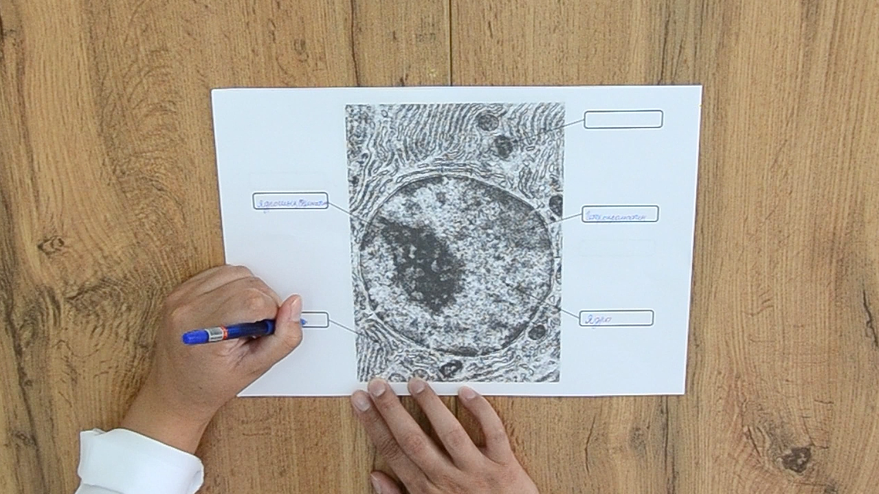
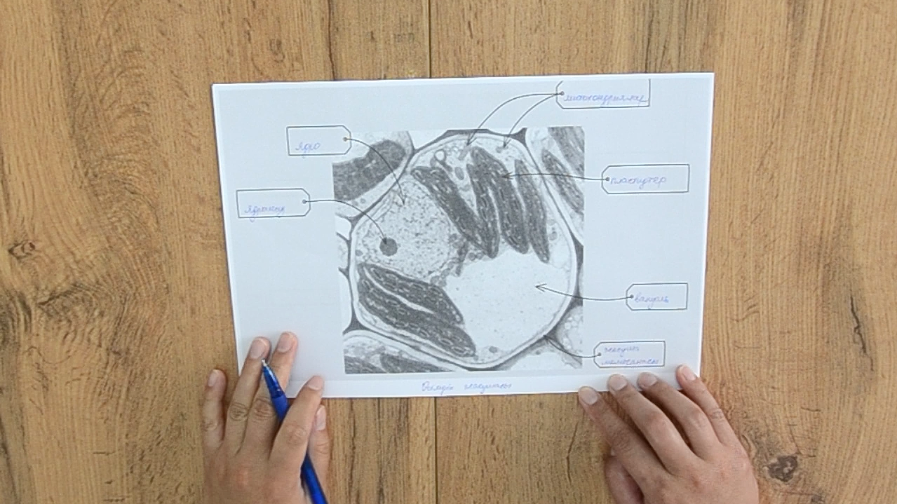
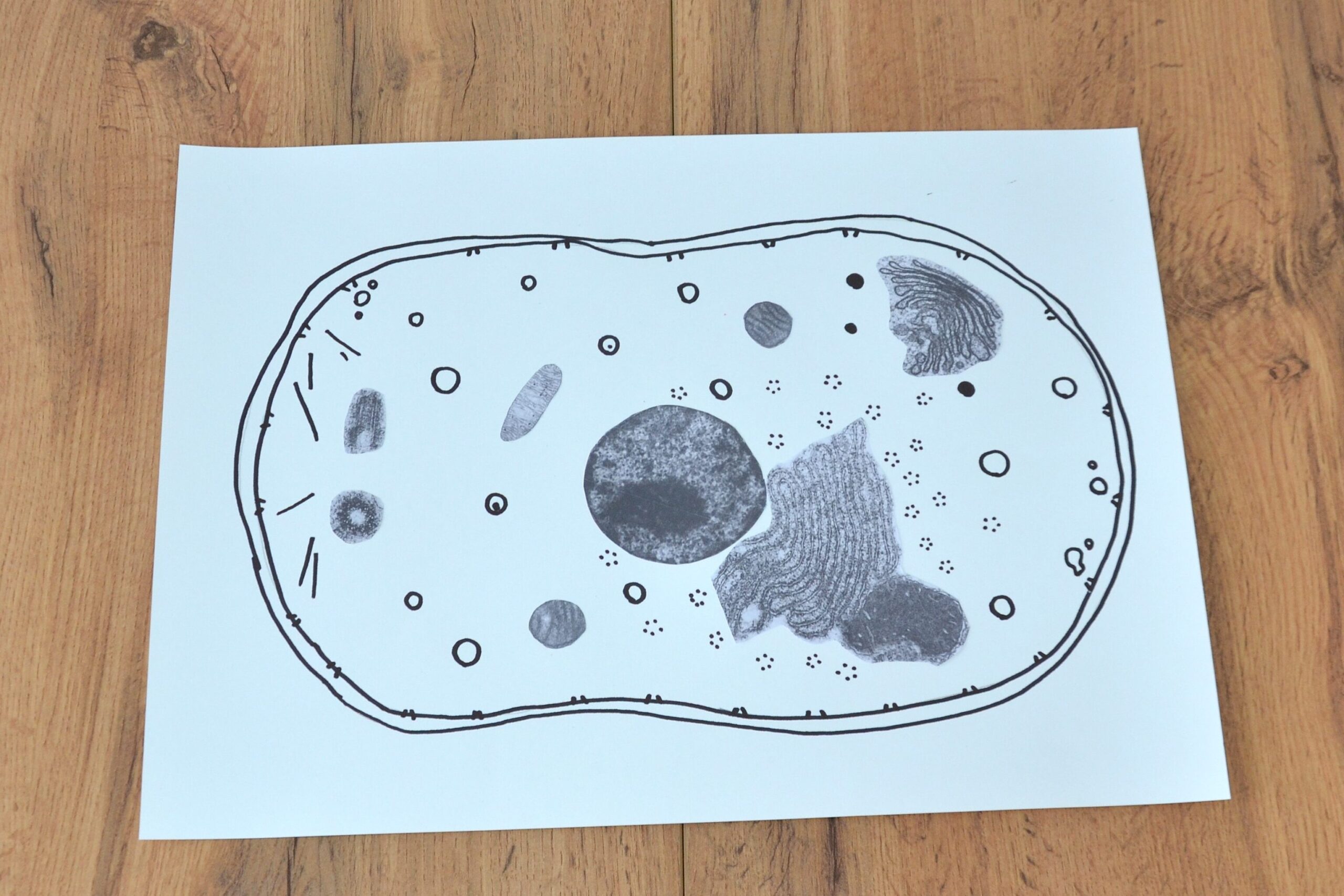
Step 3. Now observe and examine the given microphotographs. Which organelle is shown in the picture? What is its main function in the cell? Name the cellular structures located next to it.
Step 4. On an A3 sheet, draw the outline of a cell.
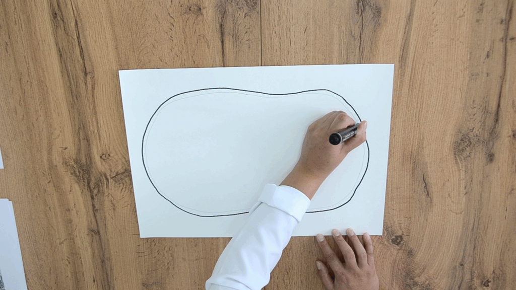
Step 5. Cut out the images of the given organelles.
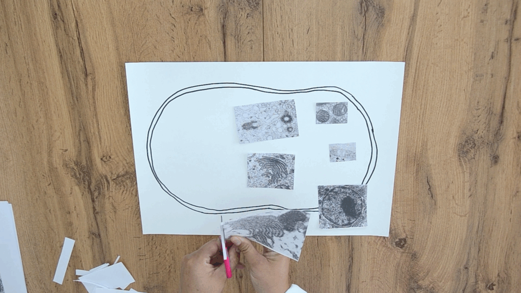
Step 6. According to your knowledge, place the organelles into the cytoplasm.
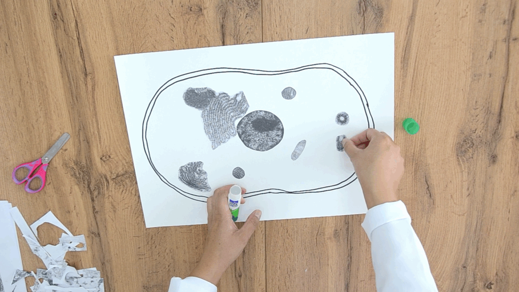
Step 7. Add small components such as ribosomes, lysosomes, vesicles, and microtubules.
Conclusion
During this work, students learned to identify the main cell organelles in microphotographs and describe their functions. The activity consolidates knowledge of cell structure, develops analytical skills, and reinforces the understanding that the cell is a complex system where all organelles function in coordination.