Lesson 1
Teacher’s Guide
– Briefly describe the topic of the lesson (introduction);
– Divide students into 3 groups;
– Monitor the execution of tasks;
– Pay special attention to homework
– Provide PBL rubrics to students to:
– the students understood in advance what criteria they need to prepare for,
– students were able to independently evaluate their colleagues.
Introduction
A person receives most of the information about the world around him through visual perception. The visual function is provided by the sensory organ – the eye. And the eyelids, eyelashes, and eyebrows protect the eyes from external damage. Tears do not allow the eyes to dry out and constantly moisten them. And also, this area of the face is actively involved in facial expressions. That is, it conveys the emotions, state, and feelings of a person well.

Let’s look at the structure of the eyes. Our eye is a complex optical system. It perceives light waves through an optical system of several layers and transmits them to a specific part of the brain.
The eyeball consists of several shells and is in the form of a sphere. Fibrous membrane – the outer shell consisting of the sclera and performing the functions of protection and shaping. The second shell – the choroid contains many vessels responsible for the blood supply and nutrition of the eyeball. The inner shell is the retina.
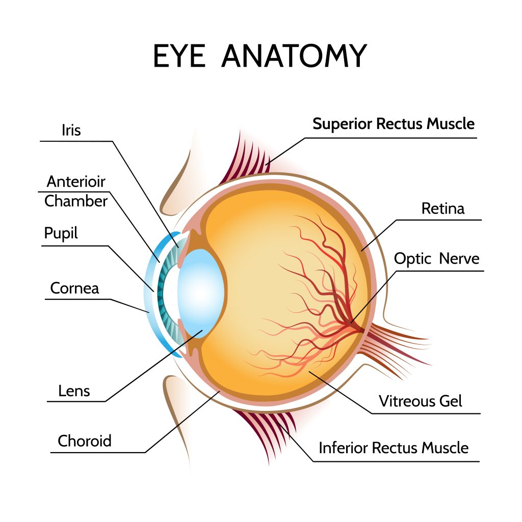
The optical system of the eyes consists of reflective lenses that form an image of the outside world on the retina. It consists of the cornea, lens, and vitreous gel, behind which is the retina, which perceives light.
Light waves entering the eye pass through the cornea – a transparent shell covering the front of the eye, which is a dome-shaped lens. It borders on the outer shell of the eye – the sclera.
Behind the cornea is the iris. The iris is a circle with a round hole inside – the pupil. The iris enters the choroid of the eye. It can change the diameter of the pupil, with the contraction and relaxation of the muscles, in response to the amount of light entering the eye. Thus, the eye adapts to different brightness and regulates the amount of light entering it. Light enters the retina through the pupil. And the amount of light is regulated by its size. In bright light, the pupil constricts, and the amount of light entering the eye decreases, and in low light, the pupil dilates, passing more light to the retina.
Behind the pupil is the lens – a lens that can change shape, resulting in a change in the radius of curvature. So a person can focus and see well both in the distance and near. The shape of the lens changes due to the ligaments of zin connecting to the ciliary muscles, which can contract and relax. When an object is close, the lens becomes more convex and refracts light more, and when you need to focus far away, the muscles relax and the lens becomes flatter.
Further, behind the lens is the vitreous gel – a transparent substance that occupies most of the eye and maintains the shape of the eyeball.
After the passage of light through the vitreous gel, it hits the retina. The retina consists of both nerve fibers and light-sensitive photoreceptors. It converts light energy into the electrical energy of a nerve impulse and sends it to the brain through the optic nerve. And the image is already analyzed in the brain.
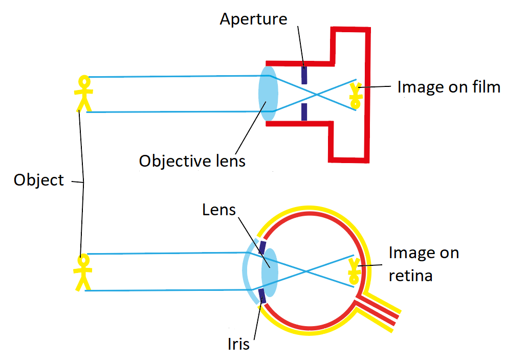
Let’s consider the eye as an optical system of lenses and compare it with a camera. They are similar in structure and function. As mentioned earlier, light rays are refracted as they pass through the cornea and lens. In the camera, this function is replaced by a camera lens. The pupil, passing light, reflexively changes the diameter and works like a diaphragm in a camera. The dimensions of the lens are changed by special muscles, changing the optical power and focusing on objects. As a result, a real, inverted, reduced image of the object is formed on the retina, as on a photographic film of a camera.
Let’s consider what problems or sore eyes can be. The science of ophthalmology deals with the study of disease and, in general, the anatomy and physiology of the eyes. There are many diseases associated with damage to the organ of vision. Some of them occur in the eye itself, in other cases, the complication of already existing diseases will involve the organs of vision in the process. Consider the most common vision problems – farsightedness and nearsightedness.
When the eyes of a person standing 5 meters from an object form an image not on the retina but in front of it, then such a visual defect is called myopia. That is, a person cannot see distant objects. When such a person squints his eyes, the pupil narrows, reducing the scattering of light rays and the eye sees a little better.
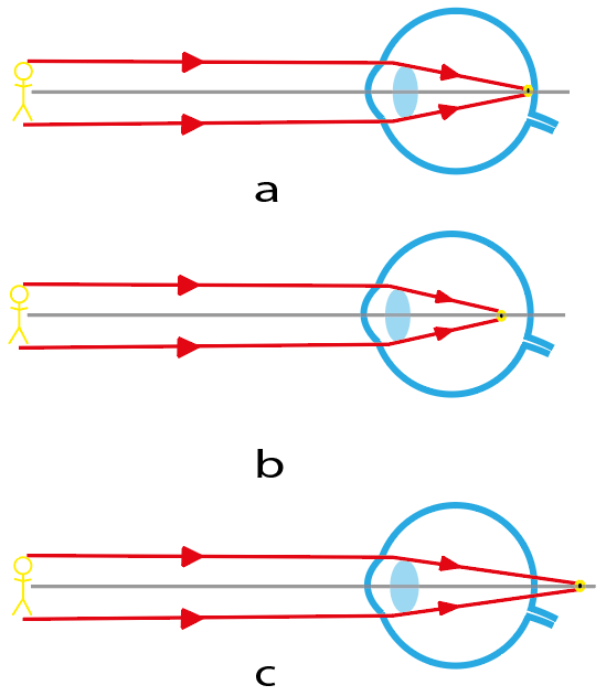 | Image of a distant object in the eye: a – normal eye; b – myopic eye; c – hyperopic eye |
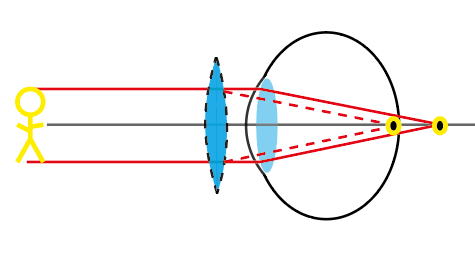
Farsightedness(hyperopia) is a visual defect in which the image of an object in the eye is formed behind the retina. At the same time, a person sees objects worse near than far away. Farsightedness is often associated with age, with time the lens becomes thinner and less elastic.
 Lenses for hyperopia | 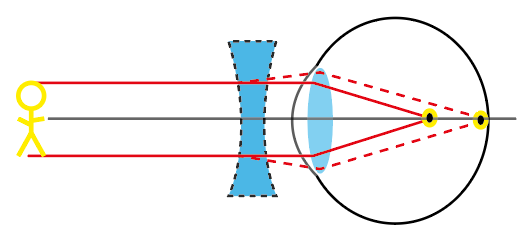 Lenses for myopia |
With the help of modern surgery, farsightedness and nearsightedness can be corrected by changing the shape of the cornea or lens. And also to correct such defects, appropriate glasses and lenses are used. A near-sighted eye needs glasses with a negative optical power, that is, diverging lenses, and a far-sighted eye need converging lenses with a positive optical power. For eyes with a defect to see normally, it is necessary to select lenses so that the image is formed on the retina.
Used media
<a href=’https://ru.freepik.com/vectors/education’>Education вектор создан(а) brgfx – ru.freepik.com</a>
<a href=”https://ru.freepik.com/vectors/education”>Education вектор создан(а) macrovector – ru.freepik.com</a>
Practical part. Vision diagnostics
Let’s determine how well our eyes see. We will check each other’s eyesight. Each group has materials for vision testing.
It is known that our eyes are exposed to high visual loads every day. This is due to prolonged work at a computer, or smartphone, relaxing in front of the TV, environmental conditions, reading in uncomfortable positions, and other factors. Therefore, regular visits to the doctor are important.
Now, we will check our vision in class and try to identify problems with vision. However, you need to understand that this will not replace a full-fledged examination and we will not be able to make diagnoses, but we will be able to identify the presence of problems and in the future will turn to doctors.
How to use the Sivtsev table to check your eyesight:
– Take the table (download) and arrange it so that the tenth line is opposite the eyes at a distance of 5 meters.
– When printing PDF files, Page Scaling must be turned off; paper size when printing = A4 (not Letter), orientation — Landscape.
– Close one eye with your hand or an opaque object (eyes should be relaxed, not closed);
– Start with letters in row 10. If you distinguish all the letters, then you have 100% vision.
– Otherwise, rise higher, to the line where you can see all the letters.
– Also check the other eye.
Visual acuity is considered complete if no more than one error was made in rows with V=0.3–0.6, and no more than two errors in rows with V>0.7. Your visual acuity score is equal to the numerical value of the letter V in the last of the lines in which you did not make mistakes beyond the norm.
Some people can distinguish letters in rows 11 and 12 from a distance of 5 meters or more without experiencing visual strain and feeling comfortable. Such people have excessively high visual acuity of 150%, and 200%. This is due to genetic characteristics.
The Golovin table works on the same principle (download). Instead of letters, it contains 12 lines with rings, each of which is torn from one side. It is necessary to determine from which side the ring is broken during testing.
Test for the definition of hyperopia and myopia
The next test is based on chromatic deviations in the eye and lies in the fact that in the optical environment of the eye, rays with shorter wavelengths (green spectrum) are refracted more strongly than longer wavelengths (red spectrum).
For test:
– Take a sheet with a table whose background is divided into green and red or open it on the screen (download).
– Move back to a distance of 70 cm – 1 m.
– If you normally wear glasses or contact lenses, you should be tested with them.
– Close one eye and try to read the letters. Check the other eye the same way.
– If you see the letters on a red background more, then it is likely that you are nearsighted; if the letters on a green background are read more clearly, then you can assume the presence of farsightedness.
– With normal vision, the eye sees letters on both sides of the same darkness and clarity.
Test for color blindness
Some people may not be aware that they misperceive a particular color. To test ourselves for volcanism, we will now take a quick and simple test. For this:
– Take Rabkin’s table. (Download)
– Relax and look at the pictures from a distance of about 1 meter.
– For each picture, you can allocate about 5-10 seconds.
– Write down what you see
– Then look at the results at the bottom of each picture.
Blind spot test
Each of us has an area on the retina that does not perceive light. It is called a blind spot. Let’s try using a simple test to find her.
– Take the paper with the test (download).
– Cover the right eye with your hand or an opaque object.
– With one eye, look at the right cross with a circle.
– Start slowly approaching and moving away from the picture without taking your eyes off the cross with the circle.
– At some point, the left cross will disappear.
– Now repeat everything with the second eye using the second sheet (download).
The fact is that in a certain place of the retina, the optic nerves gather and pass through it to connect with the brain.
And we do not notice blind spots because what one eye does not see, the second can see. Thus, our two eyes compensate for the “flaws” of each other. When we look with one eye, our brain complements the image at the site of the blind spot with what the brain thinks we should see. So, for example, during the test, instead of a cross, we saw a white background.