
Lesson
Purpose of the work:
- prepare a microscope slide from an onion root tip
- observe cell division in onion root tip cells
- explore the stages of mitosis in onion root cells using a prepared slide
Expected results:
After completing the work, students can:
- develop teamwork skills
- be able to analyze and summarize the information received
- draw logical conclusions
Teacher’s Guide:
- The task is performed in groups of 3 people
- Sharp objects should be handled with care
- Before starting laboratory work, please read the safety rules by following the link:
- To download the worksheet, follow the link:
Theoretical part
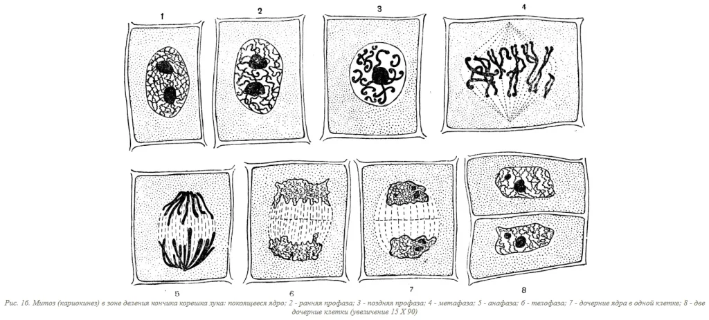
Mitosis is the process of cell division in which one cell divides into two identical daughter cells. It is the main way cells reproduce in living organisms.
Phases of mitosis
Mitosis is divided into four phases:
- Prophase. In prophase, the chromosomes condense and become visible. The centrioles move to opposite poles of the cell. The mitotic spindle begins to form.
- Metaphase. In metaphase, the chromosomes are aligned at the equator of the cell. The mitotic spindle is fully formed.
- Anaphase. In anaphase, the chromosomes separate and move to opposite poles of the cell.
- Telophase. In telophase, the chromosomes decondense. The nuclear envelope reforms. The daughter cells are formed.
Practical part
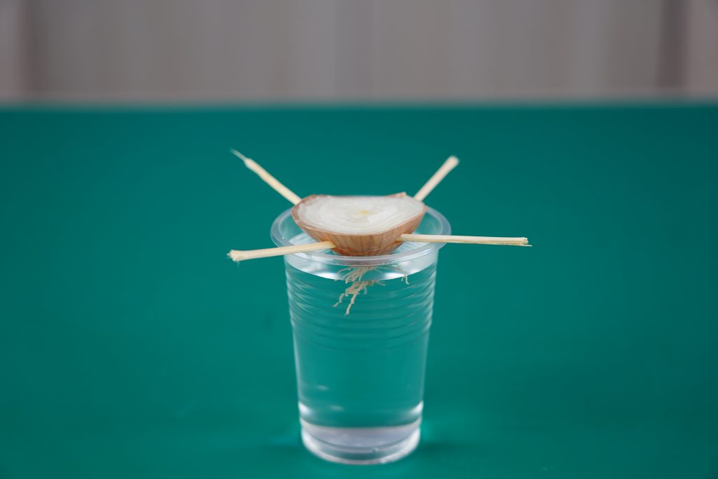
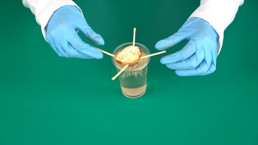
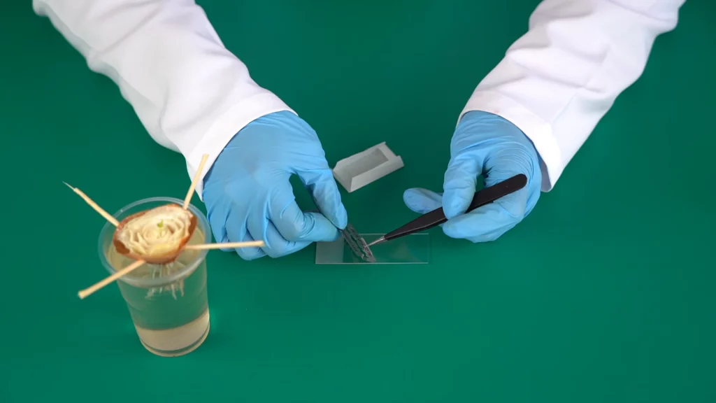
Step 1.Grow your onion roots: Place the onion in the beaker or container with its bottom submerged in water. Ensure the water level remains above the base of the onion throughout the process. Place the container in a well-lit area at room temperature and within a 2-3 days, you should see white root tips emerging from the base.
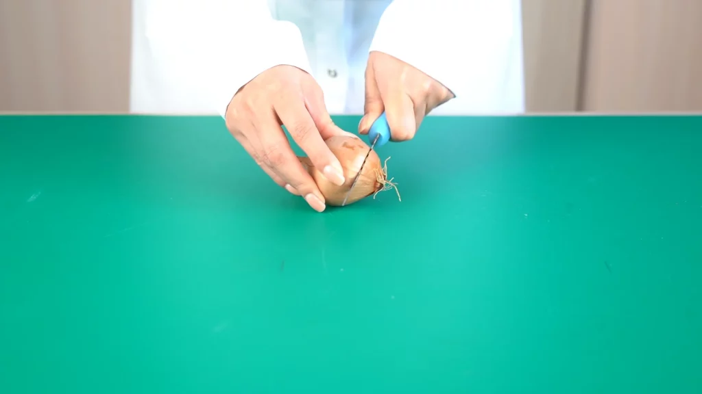
Step 2.Prepare the root tips: Choose a long, healthy root tip (around 5mm) and carefully cut it off using the sterilized scalpel or razor blade. Gently place the root tip on a clean glass slide using a tweezer.
Step 3.Fix the root tip (optional): You can fix the root tip in a solution of 3:1 ethanol to acetic acid for a few minutes to preserve its structure. This step is not always necessary, but it can help prevent cell distortion during staining.
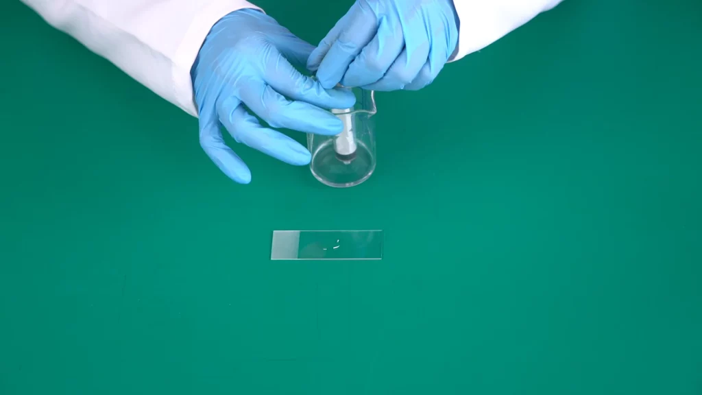
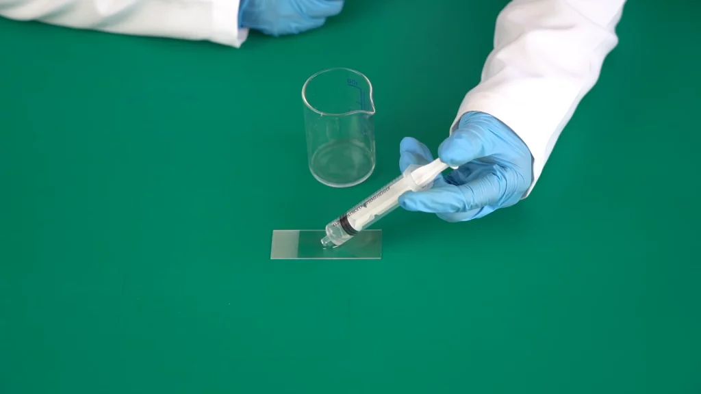
Step 4.Stain the root tip: Add a few drops of iodine or acetocarmine stain to the root tip on the slide. Gently warm the slide over a burner or hot plate (be cautious and follow safety protocols) for a few seconds to help the stain penetrate the cells. Leave the root tip in the stain for 5-10 minutes.
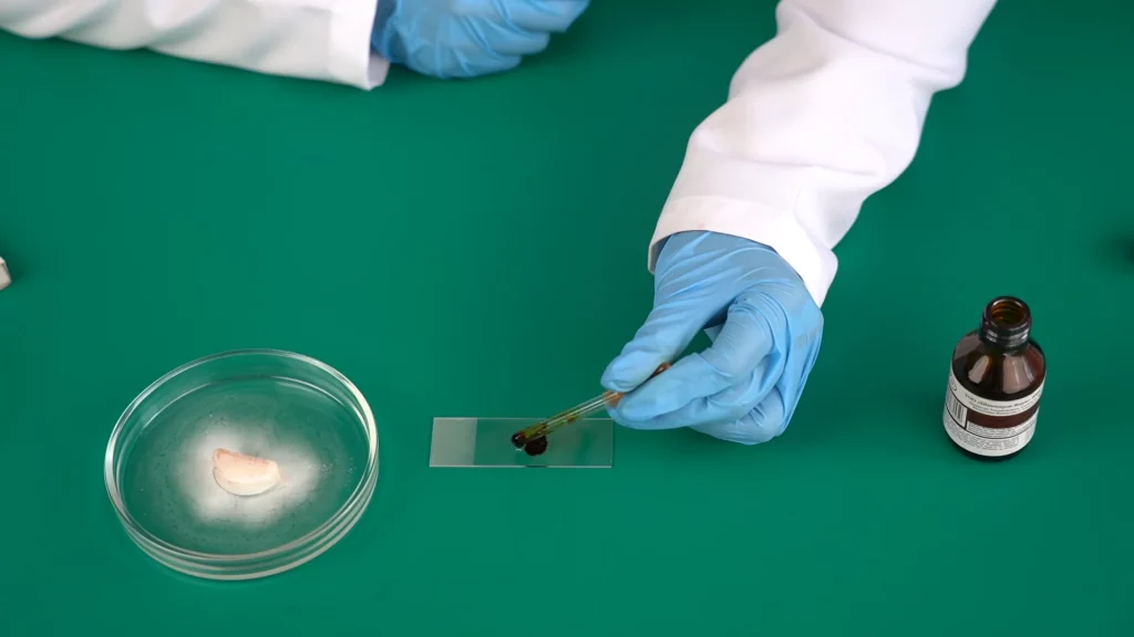
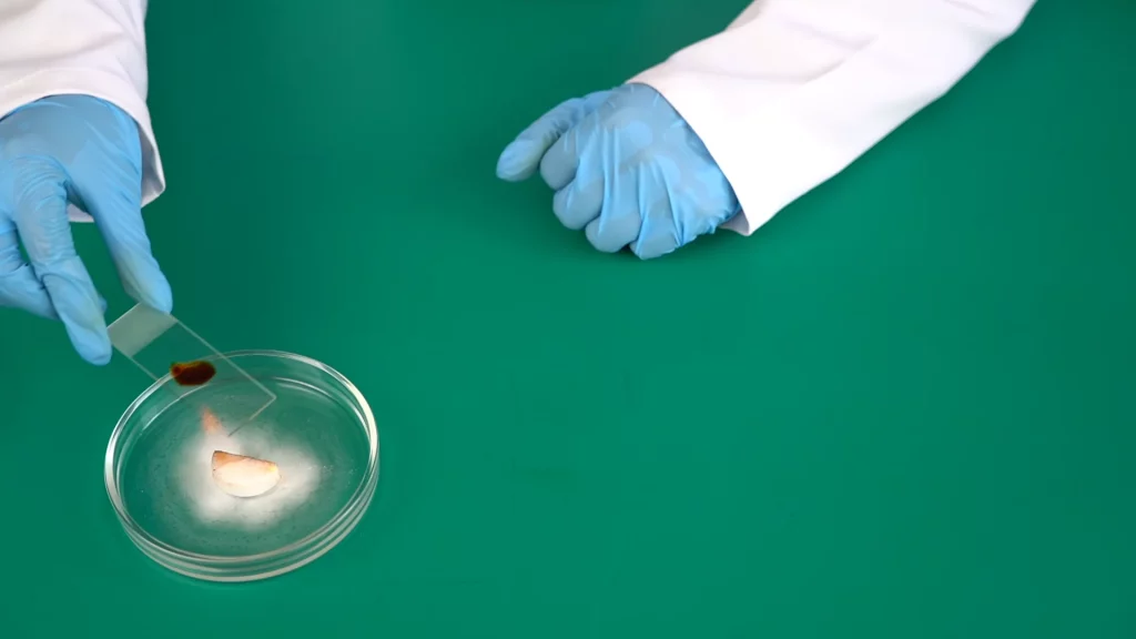
Step 5.Destain (optional): If using acetocarmine, you can briefly rinse the slide with N/10 hydrochloric acid to remove excess stain and improve chromosome visibility. Gently blot the excess liquid from the slide using filter paper.
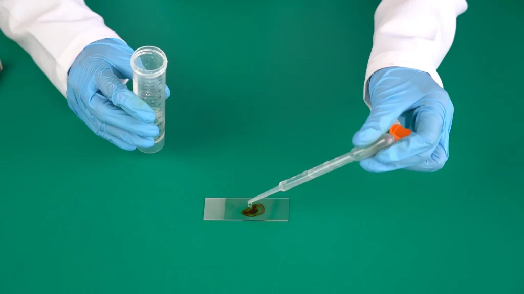
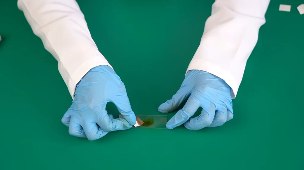
Step 6.Mount the coverslip: Add a drop of distilled water or buffer solution (depending on your staining method) to the root tip, then carefully place a coverslip over it. Avoid trapping air bubbles.
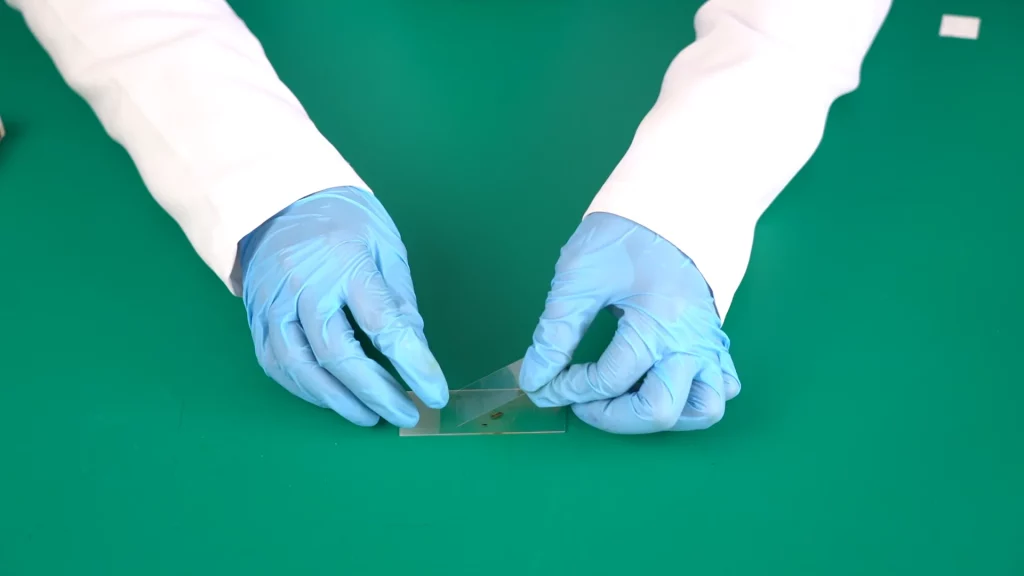
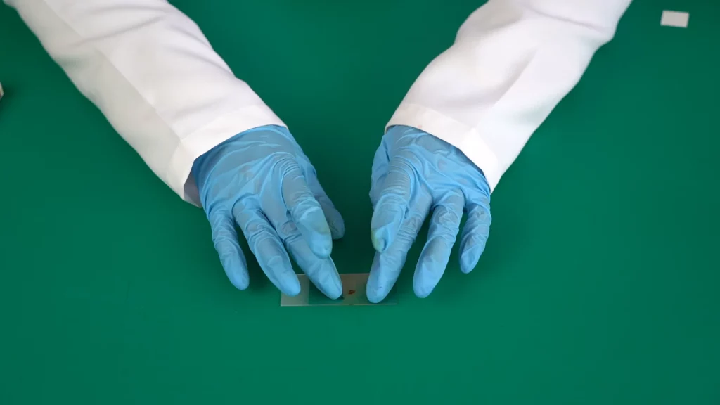
Step 7.Observe under the microscope: Adjust the microscope settings to focus on the root tip cells. You should be able to see the stained nucleus and, with luck, various stages of mitosis, including chromosomes lined up in the metaphase plate.
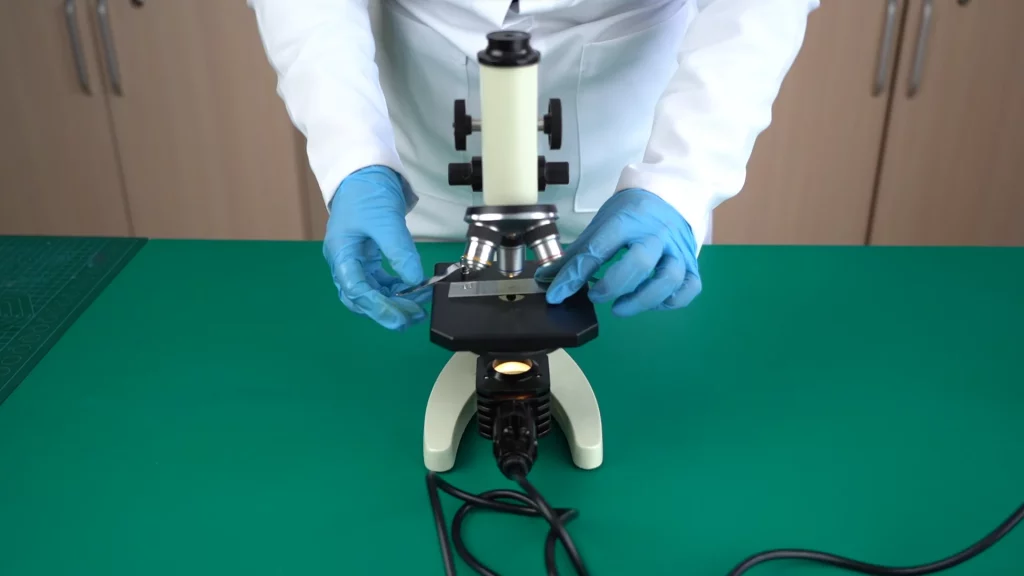
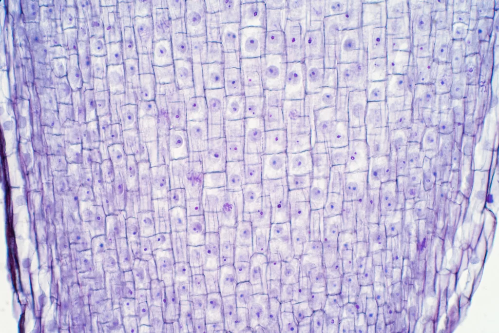
Step 8. When observing through objectives with 40x magnification, remove the coverslip from the preparation and add a couple of drops of glycerin. Then place the preparation on the stage, fix it with forceps and adjust the distance of the lens so that the nose of the lens is completely immersed in the oil.
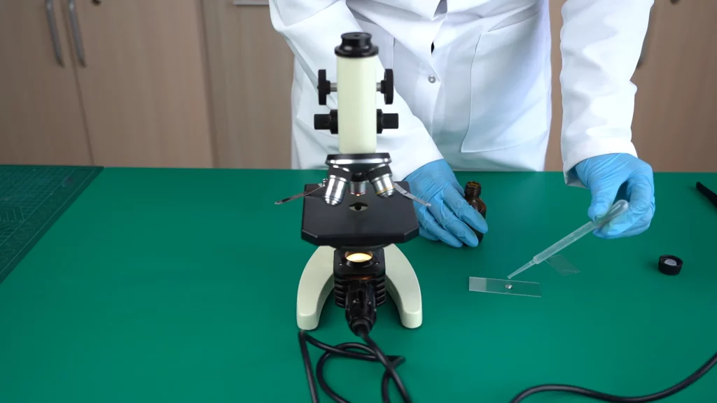
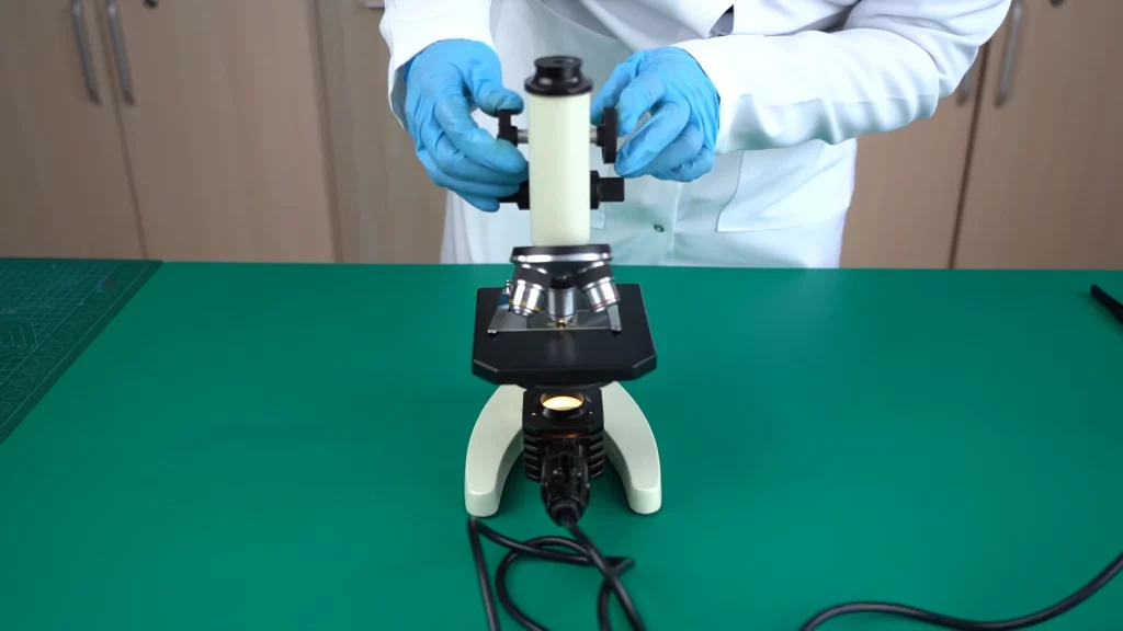
Step 9. To summarize the work, write the answers to the questions on the Worksheet.
- Why do you think the tip of the onion root was used in this experiment?
- What phases of mitosis could you see using a microscope?
- What is the biological role of mitosis in nature?
Conclusion
In conclusion, the investigation of mitosis in onion root tip slides served as a valuable learning experience, equipping students with practical skills, enhanced understanding of mitosis, and a deeper appreciation for the intricate processes that govern life at the cellular level. By utilizing prepared slides and microscopes, students gained visual confirmation of the various stages of mitosis, including prophase, metaphase, anaphase, and telophase. Through manipulating slides, adjusting microscope settings, and identifying cell structures, students refined their practical skills in microscopic observation.