
Lesson 1
Learning objectives:
- Understand the basic principles of bacterial growth.
- Practice aseptic techniques for culturing bacteria.
- Observe and analyze bacterial colonies.
- Draw conclusions based on experimental data.
Safety:
- Wash your hands thoroughly before and after the experiment.
- Be careful handling hot liquids and glassware.
- Dispose of used materials properly.
Teacher’s Guide:
- The task is performed in groups of 4 people
- The culture medium can be prepared a day before the experiment.
- Working with a hot surface requires special care (hot plate, culture medium)
- Before starting laboratory work, please read the safety rules by following the link:
- To download the worksheet, follow the link:
Theoretical part
Bacteria, though invisible to the naked eye, are the most abundant and diverse organisms on Earth. This incredible diversity extends to their shapes and characteristics, forming a fascinating tapestry of bacterial life.
| Bacteria forms | Description |
|---|---|
 | Cocci are spherical bacteria. They can be found individually, in pairs (diplococci), chains (streptococci), or grape-like clusters (staphylococci). Cocci are the cause of many diseases, including pneumonia and meningitis. |
 | Streptococci are cocci that are typically found in chains. They can be pathogenic, causing diseases such as strep throat, scarlet fever, and rheumatic fever. |
 | Bacilli are rod-shaped bacteria. They can be found individually, in pairs, chains, or groups. Bacilli include the causative agents of plague, cholera, and anthrax. |
 | Vibrios are slightly curved rod-shaped bacteria. They can be found individually or in pairs. Vibrios are the causative agents of cholera. |
 | Spirilla are spiral-shaped bacteria. They can be found individually or in pairs. Spirilla are the causative agents of syphilis and anthrax. |
 | Spirochetes are long, tightly coiled bacteria. They can be found individually or in pairs. Spirochetes are the causative agents of syphilis and leptospirosis. |
Practical part
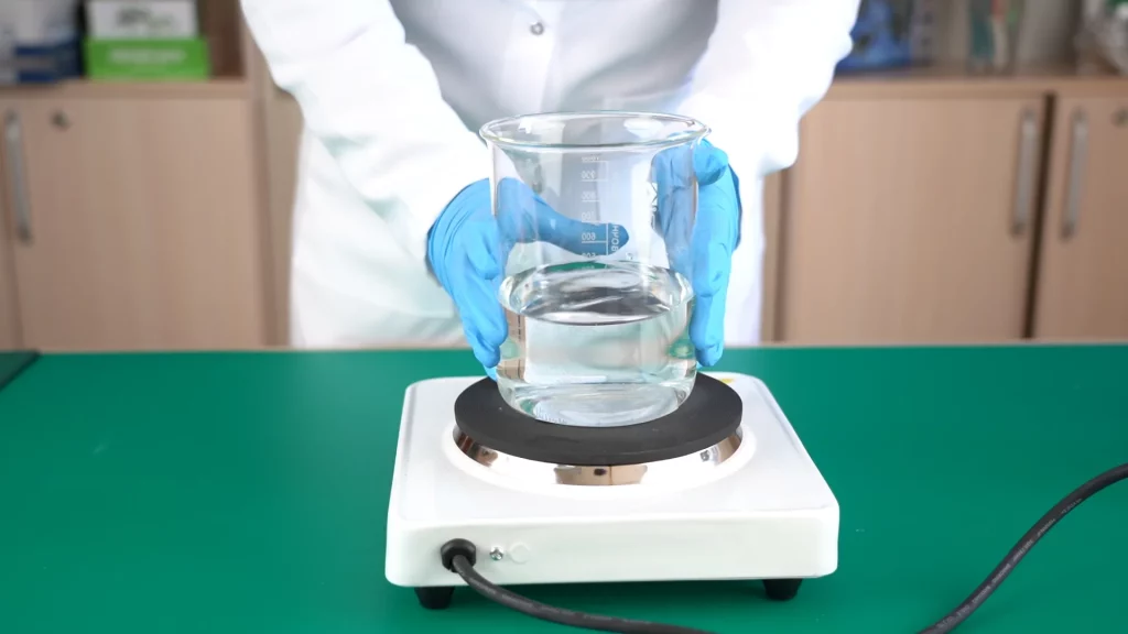
Step 1. Fill the beaker with water and place it on the stove. Boil it. Prepare a teaspoon of sugar and beef bouillon cube.
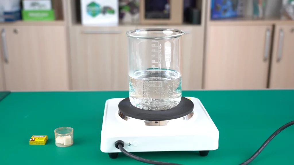
Step 2: Dissolve 1 teaspoon of sugar in it.
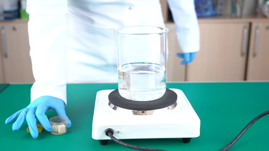
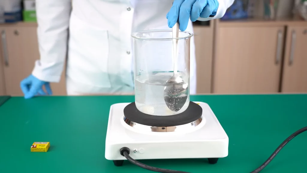
Step 3: Add a bouillon cube and dissolve it.
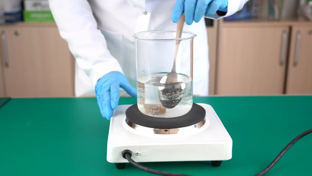
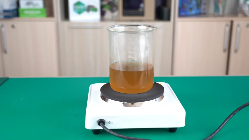
Step 4: Add agar powder and stir until fully dissolved (about 4-5 minutes)
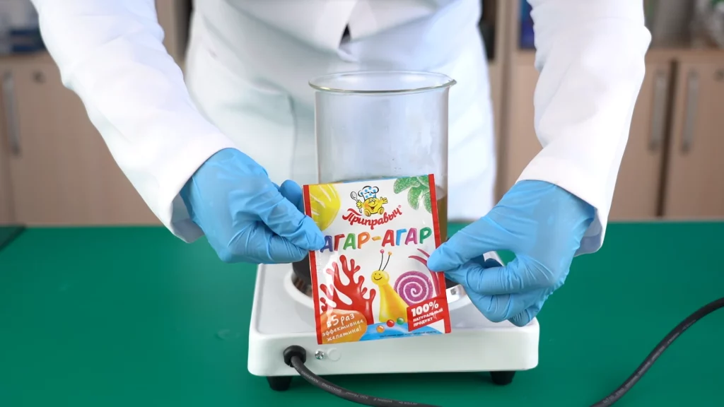
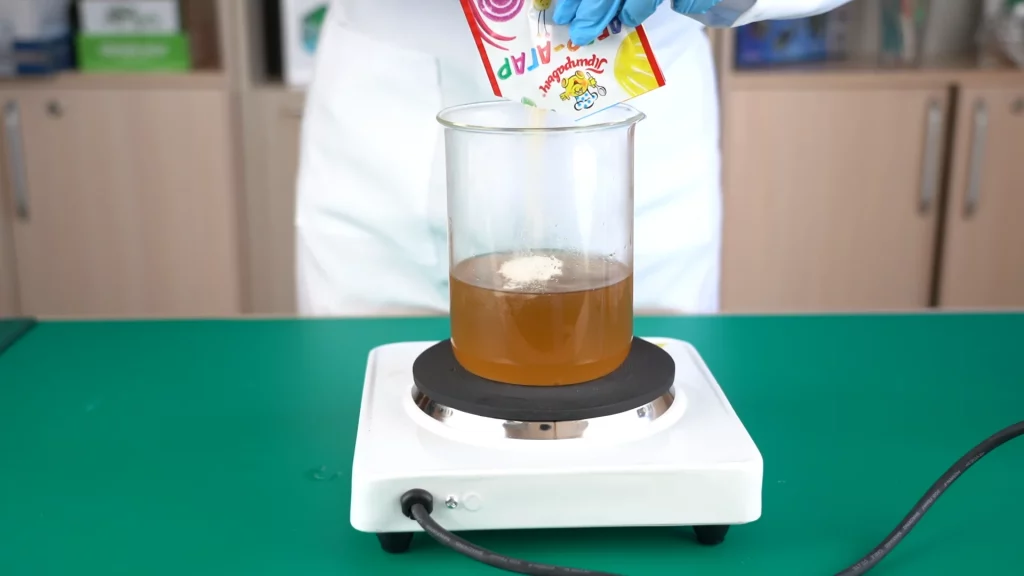
Step 5: Cool the mixture briefly. At this time, prepare the Petri dishes. Keep the lids closed until the medium is ready.
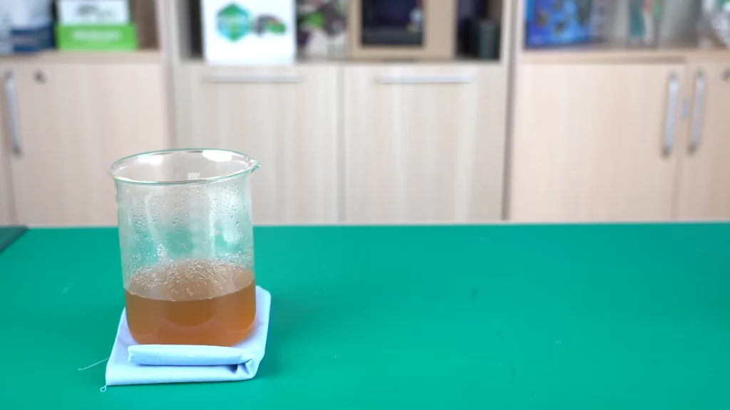
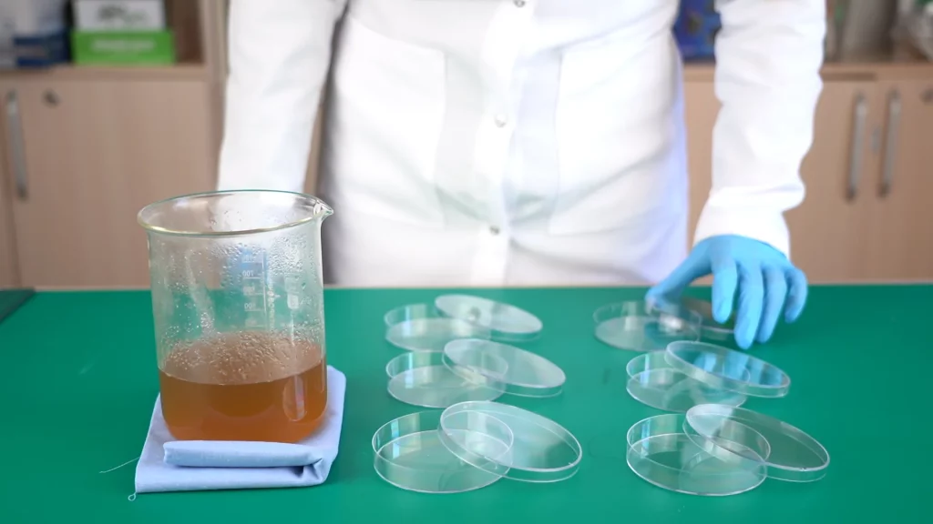
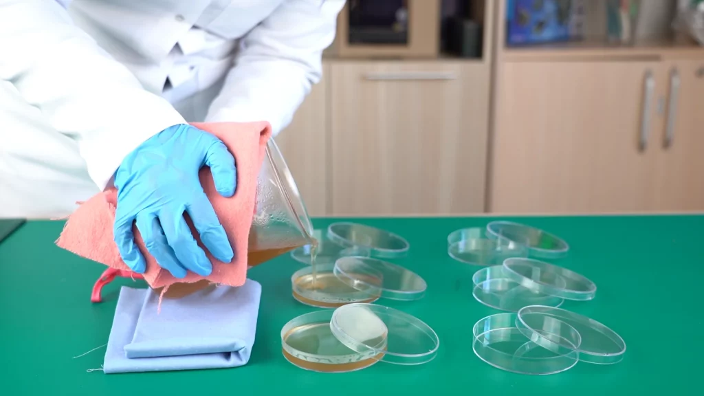
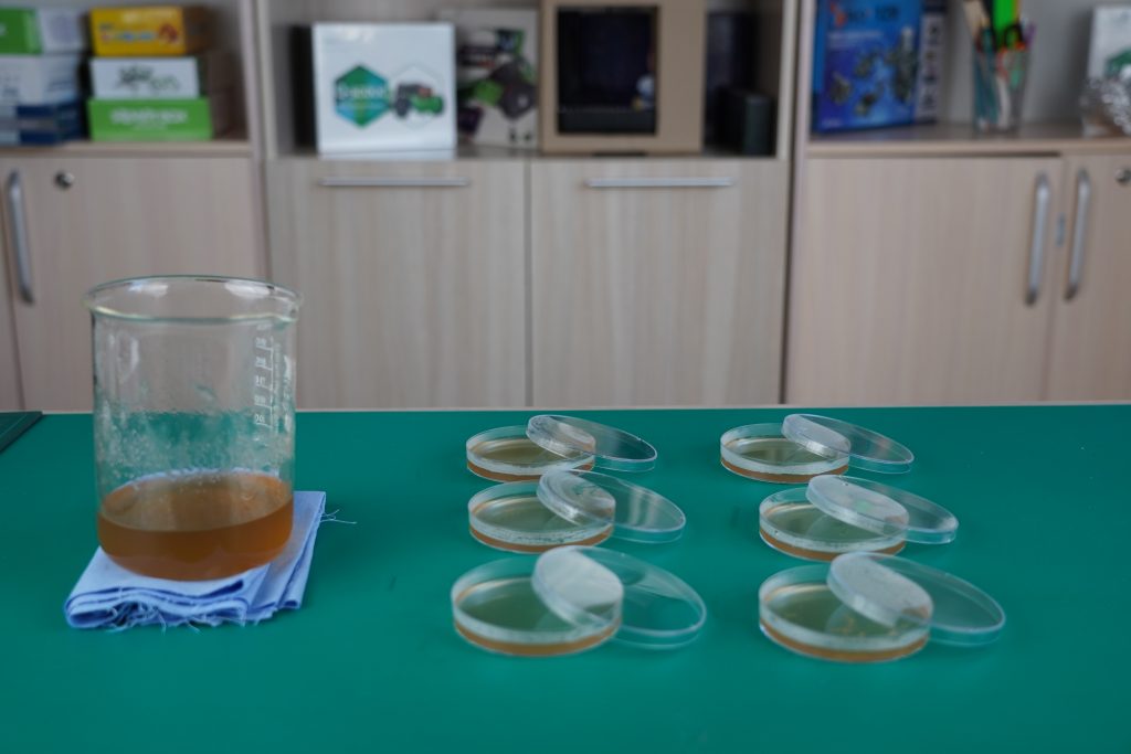
Step 6. Wear gloves and pour the prepared medium into the dishes while it’s hot to prevent premature formation of jelly in the beaker. Try to avoid unnecessary contamination, as our conditions are far from sterile. Be careful when handling hot objects. You can ask teacher to help pour the medium into the dishes. Carefully cover the cups and leave to cool at room temperature for 15-20 minutes. Allow the agar to solidify.
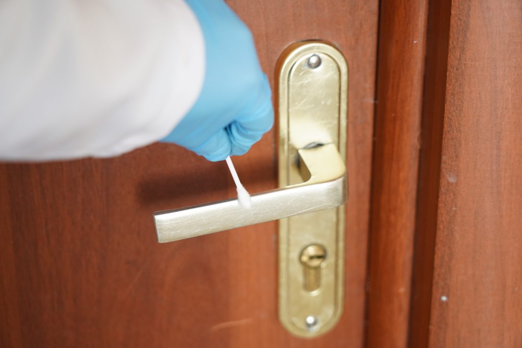
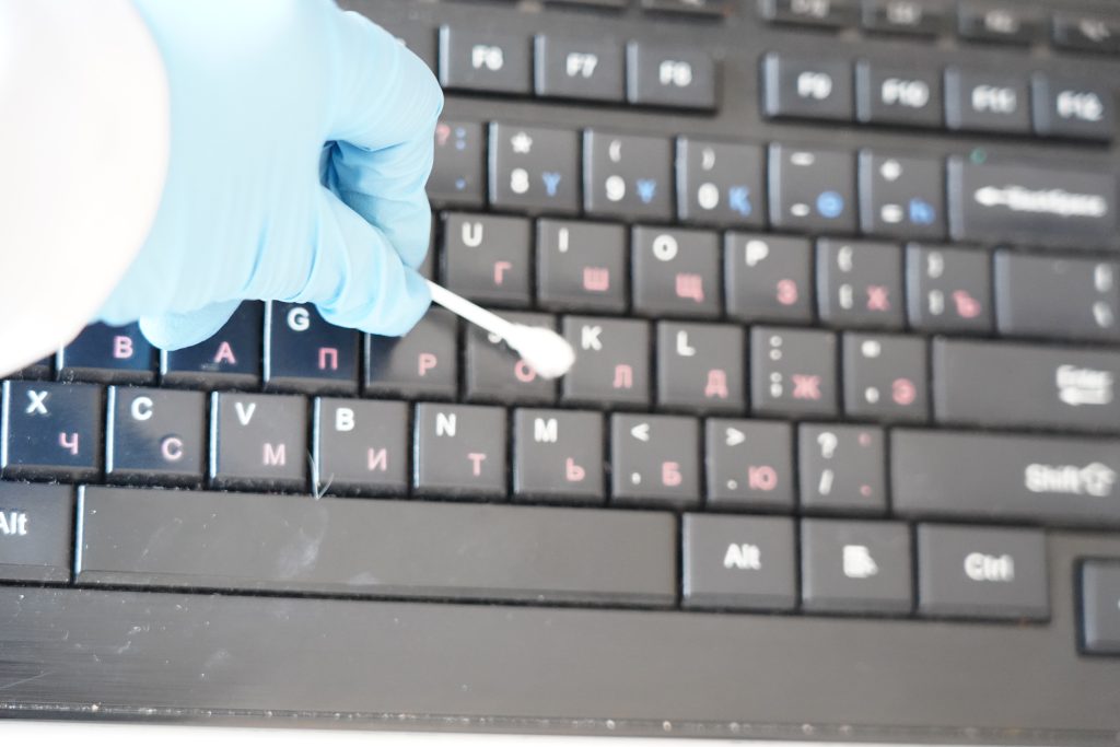
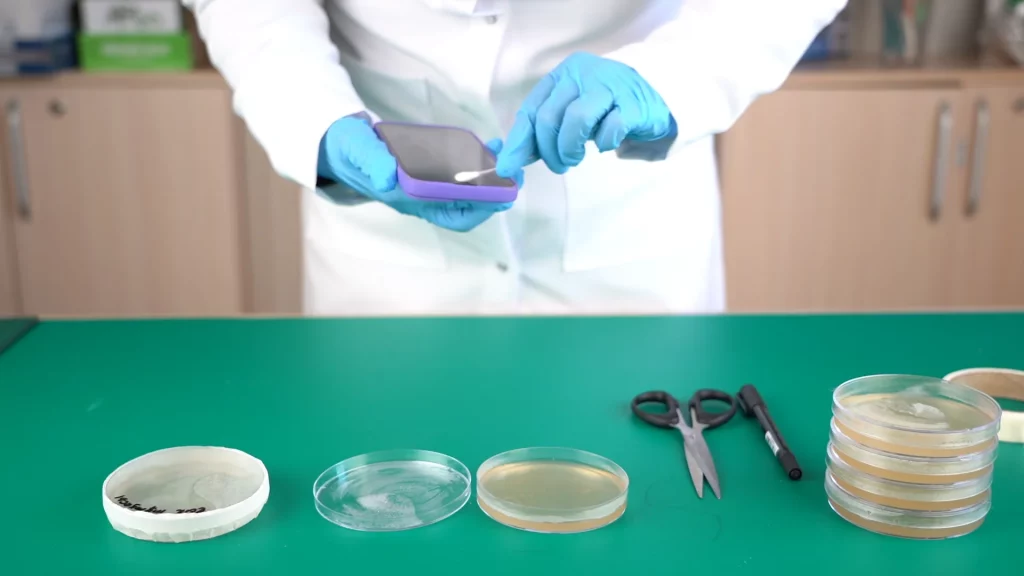
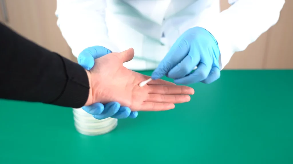
Step 7: Now it’s time to collect samples and grow colonies. Bacteria are not difficult to collect because they are everywhere.Use a clean cotton swab to gently rub surfaces you suspect harbor bacteria (hands, doorknobs, phone, keyboard).
Step 8. Gently streak the swab across the solidified agar in your Petri dish.
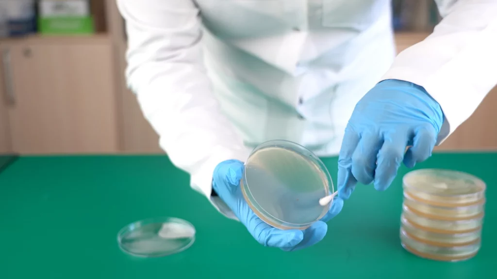

Step 9. Close the dish and secure it with plastic wrap. Label the dish with the sample source.
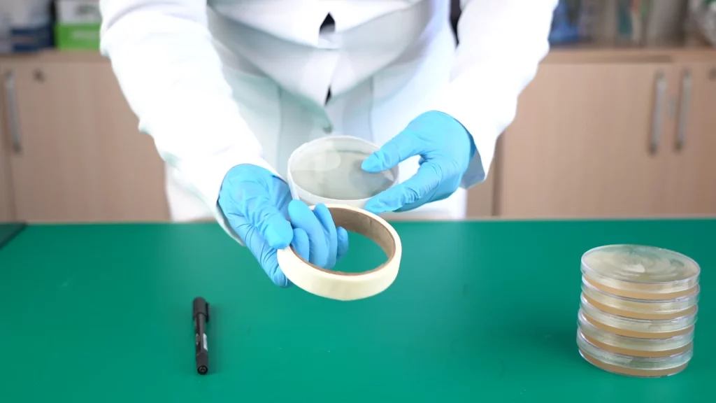
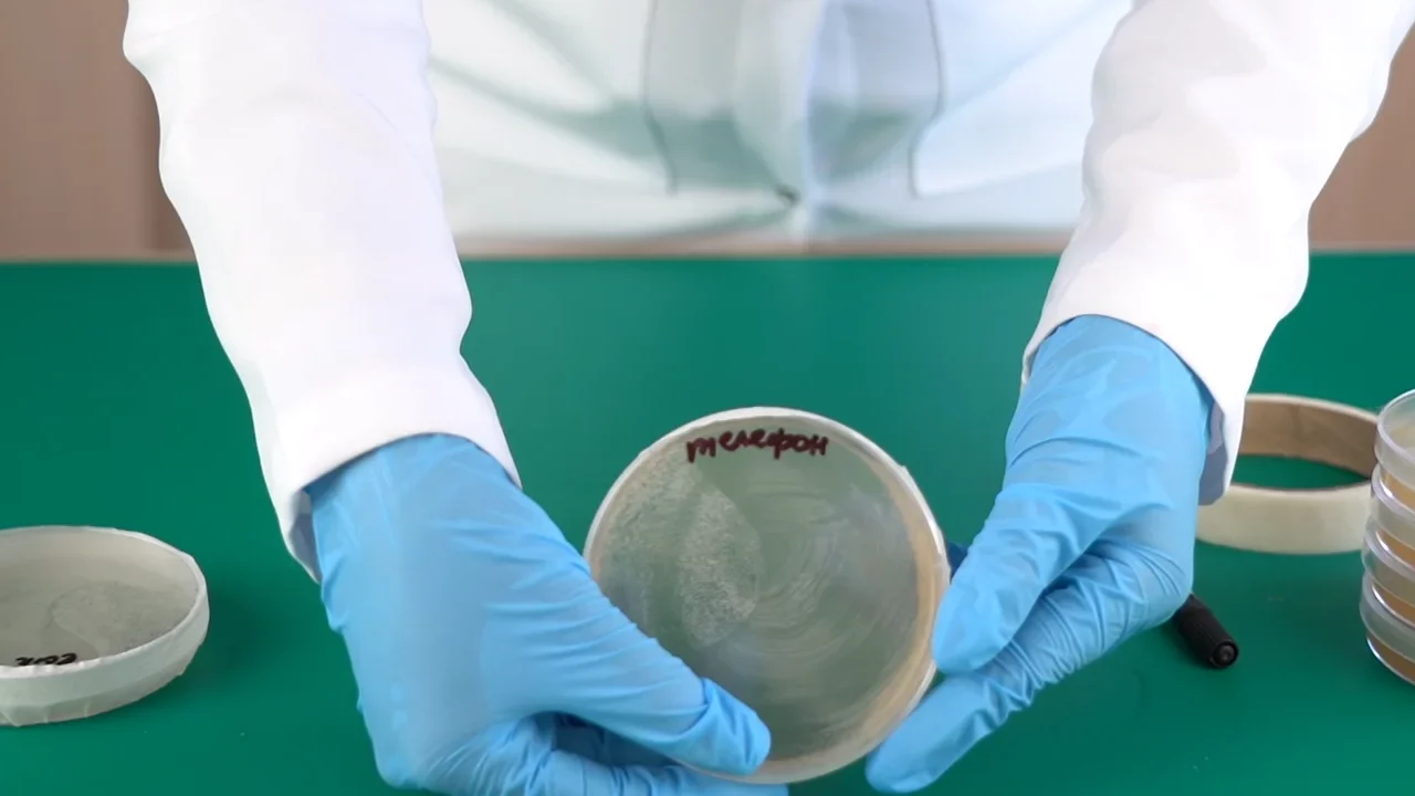
Step 10. Turn the dish upside down and place it in a warm, dark place for 48 hours. (37°C is the optimal temperature for bacterial growth). We need to turn it over so that drops that may form on the lid due to evaporation do not fall on our samples.