Week 1
Goal
1. To construct a mock-up of the human body, namely, to make a working model of the digestive system
2. Demonstrate the digestive process and study the chemical reactions occurring during digestion
3. Study the classification and composition of “fast food”
4. On the basis of experimental works to prove the harm of fast food and carbonated drinks of different origin
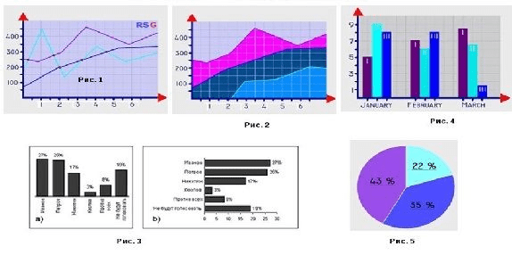
5. Conduct a survey, learn statistical data, and make a diagram based on these data.
Expected results
After studying the project, students will be able to:
– acquire the skill of cooperation with a teacher and work in a group, in pairs
– analyze and summarize the information received
– a responsible attitude to the teaching will be formed
– communicative competence will be formed in the process of educational activity
– learn how to conduct a questionnaire (survey)
– based on the survey data obtained, learn how to make graphs (charts, etc.)
– independently build logical reasoning and draw conclusions
– master the principles of proper nutrition
– to form a valuable attitude of students to their health
– study how food is processed in the human body
Interdisciplinary communication:
– Chemistry (experiments, chemical reactions)
– Biology (the human body, proper and improper nutrition, the process of digestion of food in the body)
– Mathematics (collection and analysis of information, questionnaires, statistical data)
– Artistic work (design of the layout of the human digestive system, design)
Introduction
We all know what “fast food” is, or simply, Fast Food. And many of us, at least once, have already tried such food. But few people know how much harm is caused by excessive consumption of Fast Food. And only some of us know how, under what conditions and from what Fast Food is actually prepared.
In this project, you will learn:
– classification, composition of all harmful products;
– will you conduct come chemical experiments;
– conduct a survey, study statistical data;
– and finally, evaluate where the MYTH is and where the REALITY is.
Teacher’s Guide
1. To evaluate the project, in the first week, provide this material (PBLrubrics) to students in order to:
– the students previously understood by what criteria they needed to prepare,
– the students were able to independently give an appropriate assessment to their colleagues.
2. At the beginning of the lesson, it is recommended:
– to encourage interest in the project, ask a few “leading questions”, such as:
– What depends in a person’s life on his health?
– What prevents a person from using his maximum capabilities?
– Who likes burger, cola, fries, chips, etc.?
– Who knows the composition of cola, chips, etc.?
– How many times a week do you eat Fast Food?
– Do you think “bauyrsak” refers to Fast Food?
– Is it harmful to eat Fast Food: is it a Myth or Reality? (so let’s investigate these two concepts and draw conclusions)
Description
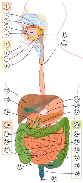
The digestive system of the human body
Diagram of the digestive tract as part of the digestive system
1. The oral cavity
2. The sky
3. Tongue
4. Language
5. Teeth
6. Salivary glands
7. Hyoid gland
8. Submandibular gland
9. Parotid gland
10. The pharynx
11. Esophagus
12. Liver
13. Gallbladder
14. Common bile duct
15. Stomach
16. Pancreas
17. Pancreatic duct
18. Small intestine
19. Duodenum
20. Jejunum
21. Ileum
22. Appendix
23. The colon
24. Transverse colon
25. Ascending colon
26. Caecum
27. Descending colon
28. Sigmoid colon
29. Rectum
30. Anal opening
| Title | Description |
| The oral cavity | The oral cavity is a bodily opening in animals and humans through which food is taken and respiration is carried out. Teeth and tongue are located in the oral cavity. In humans, it is framed by lips. In the oral cavity, mechanical grinding and processing of food by enzymes of the salivary glands takes place. The secret of the salivary glands splits the long carbohydrate chains in food into shorter ones, after which the food enters the stomach, where saliva enzymes lose their properties, since saliva enzymes can only act in an alkaline environment, and the stomach has an acidic environment. |
| The pharynx | The pharynx is part of the digestive tube and respiratory tract, which is the connecting link between the nasal cavity and mouth, on the one hand, and the esophagus and larynx, on the other. The respiratory and digestive tracts intersect in the pharynx. During swallowing, the entrance to the larynx closes the epiglottis, so food does not enter the respiratory tract, but into the esophagus. |
| The esophagus | The esophagus is a hollow muscular tube flattened in the anteroposterior direction, through which food from the pharynx enters the stomach. The motor function of the esophagus ensures the rapid advancement of the swallowed food lump into the stomach without stirring and pushing. |
| The stomach | The stomach is a hollow muscular organ located in the left hypochondrium and epigastrium. The stomach is a reservoir for ingested food, and also performs chemical digestion of this food. |
| The liver | The liver is a vital gland of the external secretion of a person, located in the abdominal cavity (abdominal cavity) under the diaphragm and performing a large number of different physiological functions. |
| The gallbladder | The gallbladder is a reservoir for the accumulation of bile, located on the visceral surface of the liver in the fossa of the same name, its parts are the bottom, body, neck, passing into the cystic duct. |
| The pancreas | The pancreas is a large organ of the digestive system of animals and humans, having extrasecretory (exocrine) and intrasecretory (endocrine) functions. |
| The duodenum | The duodenum is the initial part of the small intestine in humans, following immediately after the pylorus of the stomach. The characteristic name is due to the fact that its length is approximately twelve fingers across. |
| The small intestine | The small intestine is a section of the human digestive tract located between the stomach and colon. The digestive process mainly takes place in the small intestine: enzymes are produced in the small intestine, which, together with enzymes produced by the pancreas and gallbladder, contribute to the splitting of food into separate components. |
| The colon | The colon is the lower, final part of the digestive tract, namely the lower part of the intestine, in which water is mainly absorbed and the formation of feces from the food gruel (chyme) is formed. |
| The appendix | The appendix is an appendage of the cecum in humans. It is a blindly ending tubular formation, the lumen of which connects to the lumen of the cecum. |
| The rectum | The rectum is the final part of the digestive tract, so named for the fact that it goes straight and has no bends. The rectum is the part of the colon to the bottom (distal) from the sigmoid colon to the anus. |
Digestion of food or what components food is broken down into in the digestive system
Digestion is a process in which the digestive system extracts important nutrients for the body and, by chemical treatment, turns undigested food into waste.
Watch the video and study the table:
https://www.msdmanuals.com/ru/%D0%B4%D0%BE%D0%BC%D0%B0/multimedia/video/digestion_ru
| Digestion of food substances and their absorption in different parts of the gastrointestinal tract | |
The oral cavity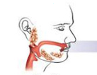 pH=5-7from slightly acidic to neutral pH=5-7from slightly acidic to neutral | Only starch begins to be partially absorbed from carbohydrates in the mouth. This is carried out by the enzyme amylase contained in the saliva. Under its influence, starch is partially broken down into small components. If you chew starchy food for a long time (which is very useful), then a small part of the starch is broken down to glucosine (a sweet taste that occurs, for example, when chewing bread). Other carbohydrates contained in food (for example, sucrose, lactose) are not broken down in the mouth. The main lipids of food are fats (triglycerides). In the mouth, they are not significantly cleaved, but still there is a sublingual lipase enzyme that cleaves a small amount of triglycerides. Digestion of proteins in the mouth does not occur. |
The stomach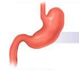 pH=1-5from strongly acidic to medium acidic pH=1-5from strongly acidic to medium acidic | The task of the stomach is to ensure mixing of the food mass coming from the esophagus and the formation of a well-mixed emulsion. Since there is a strong acidic environment in the stomach (hydrochloric acid), there is practically no further splitting of carbohydrates in the stomach. Hydrochloric acid is necessary for the coagulation of food proteins, the conversion of the enzyme pepsinogen that breaks them down into pepsin and the release of hormones that ensure the diverse work of gastric juice. Hydrochloric acid also destroys bacteria. There is an enzyme called gastric lipase in the stomach. It acts gently, but since it is relatively acid-resistant, there is still a mild cleavage of a certain amount of triglycerides. In the stomach, vitamin B12 is assimilated with the corresponding protein, which helps this vitamin to move to the place of its absorption. |
Small intestine and duodenum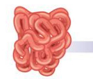 pH=6-7.5neutral pH=6-7.5neutral | In the small intestine, the food mass coming from the stomach is mixed with the enzymes of the gallbladder and pancreas. The upper part of the duodenum contains acidic gastric juice, neutral bile secret enters the lower part through the ducts of the pancreas and bile ducts. The glands in the duodenum itself produce an alkaline secretion saturated with bicarbonates. Bicarbonates and the resulting CO2 are needed to emulsify the digested food mass. B12 is released from protein and mixed for absorption with the desired protein factor. The common place of digestion of all food macronutrients (proteins, fats, carbohydrates) is the upper part of the small intestine (including the duodenum). This means that in it they are converted into smaller and simpler compounds (sugars, amino acids, fatty acids).Partial absorption of nutrients begins already in the duodenum. Here, iron and calcium are largely absorbed.By the time food enters the small intestine, called the ileum, most of the nutrients have already been absorbed. However, the importance of the ileum is primarily manifested in the fact that vitamin B12 is absorbed here, bound by the corresponding receptors. |
Large intestine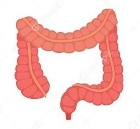 pH=5-7from slightly acidic to neutral pH=5-7from slightly acidic to neutral | A small part of the food remains undigested by the time it enters the colon. The microbiome of the digestive tract helps to split this part. |
Chemical structure of basic nutrients
| Carbohydrates | Lipids | Proteins |
Carbohydrates (sugars) are organic substances containing a carbonyl group and several hydroxyl groups. By the formula Cx(H2O)y, (the picture shows starch) (the picture shows starch) | Fats, also triglycerides, triacylglycerols are organic substances, products of esterification of carboxylic acids and glycerin triatomic alcohol. | Proteins (proteins, polypeptides) are high-molecular organic substances consisting of alpha-amino acids connected in a chain by a peptide bond.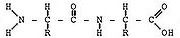 |
| The function of carbohydrates in the body:– the main source of energy in the body: 1 gram of carbohydrate = 4 kcal – enter into the composition of cells and tissues, – determine blood group, – are part of many hormones, – perform a protective function in the structure of antibodies – play the role of reserve substances in the body accumulating in the liver and muscle glycogen – temporary supply of glucose, which the body can optionally easy to use,– dietary fiber is necessary for the proper functioning of the digestive system. | The function of fats in the body:– a concentrated source of energy for the body. 1 gram of fat = 9 kcal– they participate in the processes of growth and regulation of other vital activity,– they are sources of essential polyunsaturated fatty acids,– they supply the human body with fat-soluble vitamins and are needed for their absorption and transportation in the body,– phospholipids are part of all tissues and cells, most of them are in nerve tissues and brain cells,– the fat layer formed around the organs protects them from bruises,– they are needed to remove bile into the intestines – Dietary fats are necessary because they are carriers of the flavor of food and create a feeling of satiety. Food without fat has a less pronounced taste and smell. | The function of proteins in the body:– they are necessary for the growth and construction of body cells,– almost all enzymes and some hormones have a protein composition,– actively participate in the production of antibodies and ensure the strength and activity of the immune system,– participate in the transportation of many compounds,– give food energy: 1 g = 4 kcal. |
Sources:
1. M. S. Gilyarov. Biological encyclopedic dictionary. — 1986.
2. https://toitumine.ee/ru/
3. Visual physiology / S. Zilbernagl, A. Despopoulos / Translated from English by A. S. Belyakova, A. A. Sinyushina | Moscow | BINOM. Laboratory of Knowledge
4. N. A. Abakumova, N. N. Bykova. 9. Carbohydrates // Organic chemistry and fundamentals of biochemistry. Part 1. – Tambov: GOU VPO TSTU, 2010— – ISBN 978-5-8265-0922-7.
The practical part
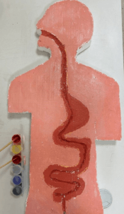
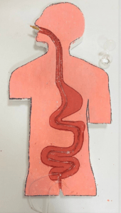
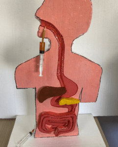
The design of the digestive system model
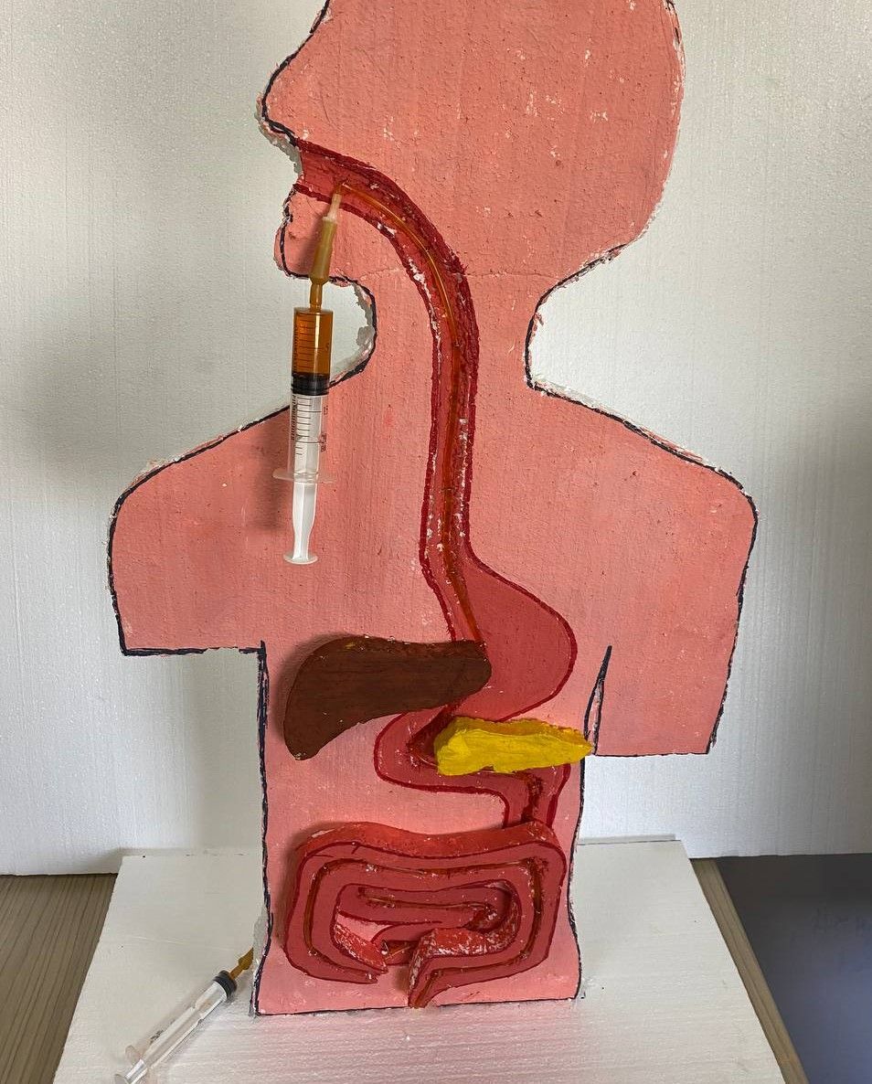
Step 2. Take a 60*80 cm foam sheet. Then draw a silhouette of the human body up to the waist.
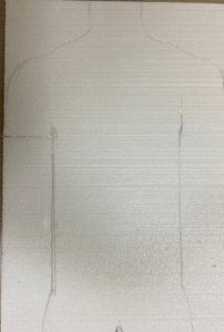
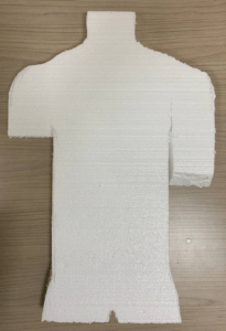
Step 3. Cut a silhouette along the line with a stationery knife.
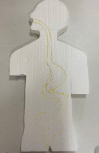
Step 4. After that, draw the digestive system.
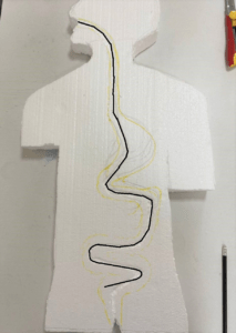
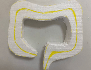
Step 5. Then cut out the pits with a stationery knife, 0.5-1 cm deep, along the line as shown in the figure
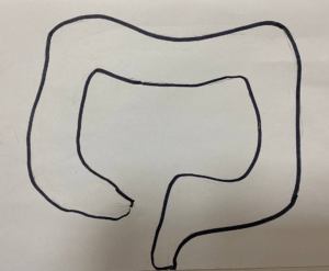
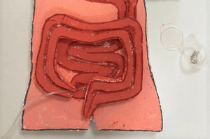
Step 6. Draw and cut out individual organs. First draw a large intestine on paper, the width should not exceed the width of the silhouette of a person. Cut out
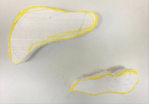
Step 7. Then take the foam of the appropriate size, and cut out the large intestine.
Step 8. In the same way, draw and cut out the liver and gallbladder and pancreas on the foam
Step 9. Painting. Mix the colors red / white in a ratio of 1/4. Then paint the human body
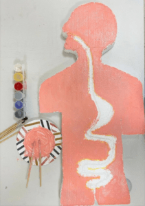
Step 10. Mix the red/white paints in a ratio of 1/2. Then color the digestive system and the large intestine
Step 11. Mix the colors red / yellow / blue in a ratio of 1/1/1 – The brown color should come out. Then color the liver
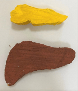
Step 12. Use yellow paint to paint the pancreas
Step 13. Use green paint to paint the gallbladder
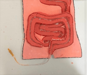
Step 14. It is necessary to install a system for infusion into the digestive system. To do this, cut out the Dropper (or filter unit). Then remove the roller clamp. And attach an infusion node from another system there. Throw the needle in the trash. All that remains is a silicone hose and infusion nodes on both sides.
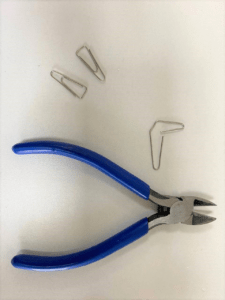
Step 15. Take paper clips, about 10 pcs. Open and divide into 2 parts.
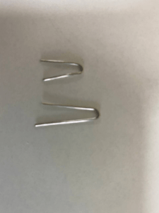
Step 16. Install the silicone hose along the cut lines. And secure the hose with pre-prepared paper clips
Step 17. After that, place it correctly outside the small intestine and glue it with hot glue.
Step 18. Then continue to stretch the system through the colon to the rectum and also fasten with paper clips.
Step 19. Prepare the solution. Pour 100 ml of water into a glass and add 0.5 tablespoons of dye.It is necessary to prepare 20 ml – 2 syringes, type the solution for one syringe.
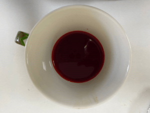
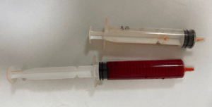
Step 20. Place the syringe with the solution in the upper part of the esophagus – in the oral cavity. And an empty syringe in another part of the infusion node.

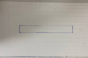
Step 21. Take a styrofoam measuring 80*40 cm

Step 22. In the middle, mark the size of the lower part of the silhouette (belt) and cut a hole about 0.5-1 cm.
Step 23. Then, insert the model into the pit using hot glue. Make sure that the model is firmly attached to the stand.
Step 24. Now check the system: Pass the solution through the system from the oral cavity, and make sure that everything is sealed. On the other hand, the syringe should open under pressure.
Homework
1. Conduct a survey during the week. (link to sample questionnaire)
2. Necessary for the next lesson (divide between groups):
– cook a cutlet (steamed / on water) from fresh minced meat (beef).
– ready burger (with beef/cutlet).
– eggshells – 4 slices
– a rusty nail or rusty coins
3. Learn the history of Fast Food
4. Learn the history of carbonated drinks
5. Build a diagram based on the received questionnaire data (the figure shows the graph templates)